Acellular Allograft Amniotic Membrane

aril® is a room temperature stable allograft derived from human placental tissue collected from consenting donors. Post-decellularization & stabilization, aril® is packaged in a double pouch packaging system and subjected to low-dose gamma irradiation. aril® is an allograft tissue intended for homologous use regulated by 21 CFR Part 1271 and Section 361 of the PHS Act.
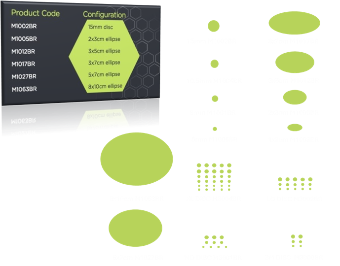
aril® is configured as a precision-cut single layer of amnion shaped as an ellipse or disc. The configuration was selected and designed to maximize clinically usable surface area and limit necessary pre-operative manipulation of the graft. Seed Biotech, Inc. has shown a greater than 95% reduction in DNA using their proprietary decellularization process. [2] As a single layer of amnion, it is not necessary to emboss aril® as there is no side-specific orientation to the graft.
Design and Handling
aril® is an acellular, precision-cut graft, designed and produced under industry-expert guidance for consistency, reliable handling, and clinical practicality.
Homologous Use
Amniotic membrane has a long, well-documented history of use as a covering and protective barrier across ophthalmic, surgical, and wound-covering settings. As a single-layer amnion allograft, aril® serves in its native function: covering the site, providing a protective biologic barrier, and acting as a scaffold. Seed Biotech® combines the natural structure of amniotic membrane with its proprietary processing to produce a consistent, acellular allograft, handled to established tissue-industry standards.
Processing Defines What Remains
Amniotic membrane allografts are not processed the same way, and the method determines what stays in the graft. Grafts preserved by cryopreservation or chemical cross-linking retain donor cells and genetic material, and some processes introduce residual agents. aril® takes a different path: decellularization. Cells, intracellular DNA, and antigens are removed while the extracellular matrix and basement membrane are preserved.
aril® is processed to greater than 95% donor DNA reduction, with residual DNA quantified lot-to-lot against defined release criteria. We report the characterization and leave the clinical judgment to the treating physician.
Available Configurations
| Product Code | Format | Size |
|---|---|---|
| M1006BR | Disc | 5 mm |
| M1001BR | Disc | 8 mm |
| M1004BR | Disc | 10.5 mm |
| M1002BR | Disc | 15 mm |
| M1003BR | Ellipse | 1 cm x 2 cm |
| M1005BR | Ellipse | 2 cm x 3 cm |
| M1012BR | Ellipse | 3 cm x 5 cm |
| M1017BR | Ellipse | 3 cm x 7 cm |
| M1027BR | Ellipse | 5 cm x 7 cm |
| M1063BR | Ellipse | 8 cm x 10 cm |
Start a Conversation
Amniotic Membrane Origins
The amniotic membrane, the innermost layer of the placenta, has a long history of surgical use across multiple disciplines, and a correspondingly deep record of its handling and safety. After thorough screening, eligible mothers can consent to donate this tissue via a scheduled Cesarean-section birth. This information is intended to inform medical professionals, researchers, and individuals involved in tissue donation and transplantation.[1]
Ocular Surface Use
aril®, an acellular allograft amniotic membrane, is used as a covering and protective barrier on the ocular surface across a range of surgical and ocular-surface settings. Product format, sizing, and placement are determined by the treating clinician.
Surgical Use
aril®, an acellular allograft amniotic membrane, is used as a covering, protective barrier, and structural scaffold across reconstructive and gynecologic surgical settings. Product format, sizing, and placement are determined by the treating surgeon.
Tissue Characteristics
Acellular, greater than 95% DNA reduction
Single-layer amnion
Precision-cut disc or ellipse
Room-temperature stable
Terminally processed by low-dose gamma irradiation
No side-specific orientation

aril® for the ocular surface
aril® IFU
1 Davis JS (1910) Skin transplantation with a review of 550 cases at the Johns Hopkins Hospital.
2 Early, Ryanne. Decellularized biomaterial from non-mammalian tissue. WO 2014110269, Jul 17, 2014A1